
Privacy statement: Your privacy is very important to Us. Our company promises not to disclose your personal information to any external company with out your explicit permission.
Introduction
Laser and non-laser light sources for hair removal are one of the most common and popular cosmetic procedures. The different lasers and light sources currently available for photoepilation operate in the red or near-infrared wavelengths and include ruby laser (694nm), alexandrite laser (755nm), diode laser (800 – 810nm), Nd:YAG laser (1064nm), and Intense Pulsed Light (IPL) (590 – 1200nm) these are Professional Aesthetic Equipment. . All of the lasers and light sources are based on the principle of selective photothermolysis. This principle states that through the use of lasers or light sources, a thermal injury can be confined to a specific chromophore within the skin and to a specific target, but other structures with different chromophores are left untouched. Laser hair removal is mainly based on the specific targeting of melanin in the hair bulb. Melanin absorbs the light emitted by the laser at a specific wavelength. The energy of the laser is converted into heat and, thus, causes the selective destruction of the hair bulb.
However, melanin in the surrounding epidermis can also be targeted. Therefore, presently, complete selective damage during laser treatments is not possible. The adverse effects of laser hair removal such as blistering, pigmentary changes and scarring are primarily related to this unwanted epidermal damage after partial absorption of the laser energy. Despite the development of various strategies such as cooling systems, laser procedures still present a risk of adverse effects due to the overheating of skin.
Diode lasers are solid-state laser devices that have been successfully used over the past several years. Because of their reliability and their ability to penetrate into the much deeper part of the skin, even darker-skinned individuals are successfully treated for the epilation of unwanted hair. Clinical studies using diode lasers have shown their effectiveness in permanent (long-term) hair removal and have had minimal adverse effects.
Our study was designed to investigate the effects of 810nm diode laser treatment on hair and on the biophysical properties of the skin by using various noninvasive techniques on various parameters, including hair analysis, surface color changes, the integrity of the skin barrier, sebum production rate and pH level.
Materials and methods
Subjects
Thirty-five women with at least moderately dense, dark, axillary hair were enrolled in our study between March 2007 and June 2008. Exclusion criteria were as follows: age below 18 years, pregnant or breastfeeding women, any hormonal dysfunction, use of medications or hormones with androgenic effects, a history of recent tanning, hypertrophic scars, keloids, photosensitivity or those taking medications known to cause photosensitivity, any infection or inflammation in the laser area, any previous electrolysis and laser treatment to the study areas, waxing, depilation or bleach use within 2 months of entry into the study, shaving or clipping of hair within 2 weeks of entry into the study, isotretinoin use within 6 months of entry into the study, and seizure disorder triggered by infrared light. Using topicals or cosmetics were strictly avoided during the study in order that any kind of applications should not produce changes in barrier function.
Laser procedure
In order to determine whether laser therapy would be performed on the left or right side, randomization was conducted by drawing. The diode laser at an 810nm wavelength with variable pulse duration up to 100 ms was used in our study. Prior to the treatment, hairs were shaved with a safety razor. Immediately before laser treatment, a transparent gel was applied to the skin surface. The laser was used in the PRO1 and BASIC-mode with a 12-mm spot size and radiant exposure 25 – 30 J/cm2 . The epidermis was cooled immediately before laser irradiation by placing a cold aluminum applicator plate onto the skin surface.
Hair analysis
Hair analysis was performed before the laser treatment and at weeks 2, 4 and 6 after laser therapy. The measurements were made from the center of the mid-axillary line after the biophysical measurements. While measuring hair density and hair thickness, we made use of the digital epiluminescence microscopy (ELM) system and TrichoScan Professional Edition software. However, since TrichoScan Professional Edition software was specifically developed to analyze scalp hair, a visual hair assessment method was used. Digital images were obtained at 30-fold (analyzed area 0.586 cm2 ) magnification with the digital ELM system.
Table I. The results of hair analysis.

In order to determine hair densities, we first visually counted the hairs in the target area photographed with x 30 enlargements and then divided the hairs counted by area. We measured hair thicknesses by transversely marking each hair in the target area photographed with x 30 enlargements from a distance of 1 mm from the proximal portion. Then, the average hair thickness was determined.
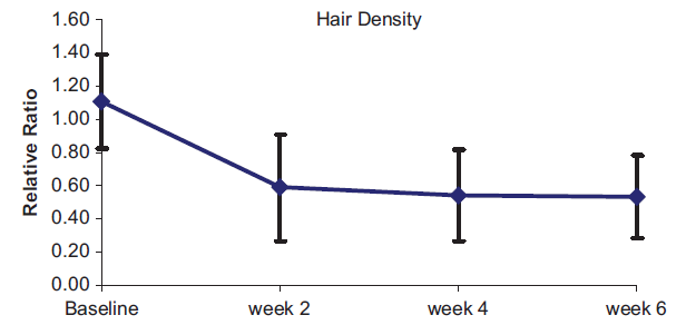
Figure 1. Changes of hair density over time. Data are shown as mean ± SD and are expressed as the ratios of the laser-treated areas to the control areas. Hair density statistically significantly decreased in the first evaluation, and the difference remained during the subsequent measurement.
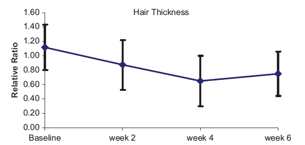
Figure 2. Changes of hair thickness over time. Data are shown as mean ± SD and are expressed as the ratios of the laser-treated areas to the control areas. Hair thicknesses statistically significantly decreased in the first evaluation and the difference remained during the subsequent measurement.
Biophysical measurements with non-invasive devices
Biophysical properties were assessed by using different non-invasive techniques on various parameters before the treatment and at weeks 2, 4 and 6 after the therapy. In all the measurements, except for the sebumeter, two consecutive readings were taken at each site and their mean values were calculated. The center of the mid-axillary line was considered as the guide and the measurements from the periphery of the guide were conducted in the way that only one measurement probe could touch one single area. Measurements were made at normal room conditions (20℃ and 40 – 60% air humidity). Subjects were required not to do any physical activity within 30 minutes before the measurement and were acclimatized for at least 30 minutes to the standard room conditions. Measurements were obtained between 2:00 pm and 3:00 pm due to diurnal variations.
Skin reflectance. Erythema and skin pigmentation were measured based on the absorption/reflexion. The melanin index (MI) and erythema index (EI) were calculated at each visit. The Mexameter shows that the melanin and erythema values range from 0 to 999.
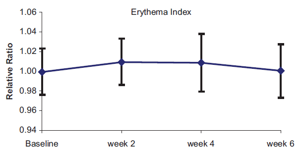
Figure 3. Changes of erythema index over time. Data are shown as mean ± SD and are expressed as the ratios of the laser-treated areas to the control areas. A statistically significant increased erythema index was only observed in the first evaluation at 2 weeks.
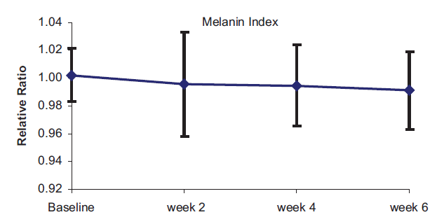
Figure 4. Changes of melanin index over time. Data are shown as mean ± SD and are expressed as the ratios of the laser-treated areas to the control areas. There was no statistically significant difference between the areas.
Transepidermal water loss. Transepidermal water loss was measured based on the diffusion law. The measuring range of the device is 0 – 320 and all the values were expressed in g/m2 per hour.
Capacity of stratum corneum hydration. The water-holding capacity was measured based on capacitance measurement of dielectric medium. The measuring range of the device is 0 – 130 and all the values were expressed in arbitrary units.
Sebum analysis. The amount of sebum secretion was measured based on grease-spot photometry. The measuring range of the device is 0 – 350, and all the values were expressed in μg/cm2 .
pH analysis. Measurement of the pH value covers an important characteristic of any aqueous solution: its acidity or alkalinity. The measuring range of the device is from 0 to 12.
Statistical analysis
Parametric methods were applied in our study. The relationship between the values of pretreatment and the other three measured values obtained during the 6-week period was analyzed statistically. A p -value 0.05 was regarded as statistically significant. Differences between the laser-treated areas and the control area at pretreatment and during the 6-week follow-up period were analyzed using the paired samples t -test for dependent variables. The Bonferroni correction was used to make statistical adjustments for multiple comparisons, and a p -value 0.008 was considered to be
statistically significant.
Results
Thirty-one women between 18 and 45 years of age completed the study. A total of four subjects were lost at different times during the follow-up period of the study. Of the subjects, four had Fitzpatrick ` s skin type II, 22 had type III and five had type IV.
Hair analysis
The results of hair analysis are shown in Table I. The laser-treated areas showed significant hair loss compared with baseline. Hair density statistically significantly decreased in the first evaluation conducted 2 weeks after the laser treatment and the
statistically significant difference remained during subsequent measurements (Figure 1). Similarly, hair thicknesses were significantly smaller after the laser treatment. Hair thicknesses statistically significantly decreased in the first evaluation conducted 2 weeks after the laser treatment and the statistically significant difference remained during subsequent measurements (Figure 2).
Biophysical measurements with non-invasive devices
Skin reflectance. When the EI in the laser-treated areas was compared with the EI in the control areas, there was a statistically significant increase in the laser treated areas in the first evaluation at 2 weeks. In subsequent evaluations, there were no statistically significant differences (Figure 3). In the MI, there were no statistically significant changes during the measurement period (Figure 4).
Transepidermal water loss. When the evaluations of Tewameter scores of the treated and untreated areas during the measurement period were examined, there was no statistically significant difference between them (Figure 5).
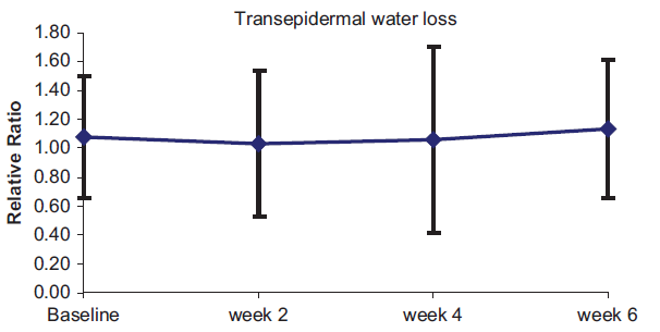
Figure 5. Changes of transepidermal water loss over time. Data are shown as mean ± SD and are expressed as the ratios of the laser-treated areas to the control areas. There was no statistically significant difference between the areas.
Capacity of stratum corneum hydration. When the evaluations of Corneometer scores of the treated and untreated areas during the measurement period were examined, there was no statistically significant difference between them (Figure 6).
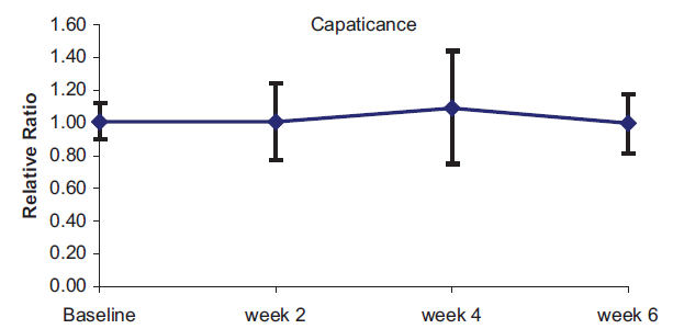
Figure 6. Changes of capacity of stratum corneum hydration over time. Data are shown as mean ± SD and are expressed as the ratios of the laser-treated areas to the control areas. There was no statistically significant difference between the areas.
Sebum analysis. When the evaluations of Sebumeter scores of the treated and untreated areas during the measurement period were examined, there was no statistically significant difference between them (Figure 7).
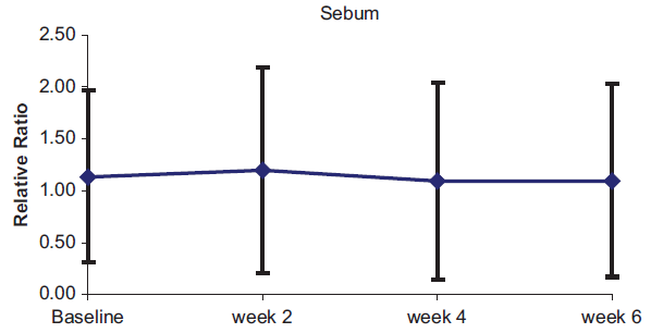
Figure 7. Changes of sebum secretion over time. Data are shown as mean ± SD and are expressed as the ratios of the laser-treated areas to the control areas. There was no statistically significant difference between the areas.
pH analysis. When the evaluations of Skin-pH-Meter scores of the treated and untreated areas during the measurement period were examined, there was no statistically significant difference between them (Figure 8).
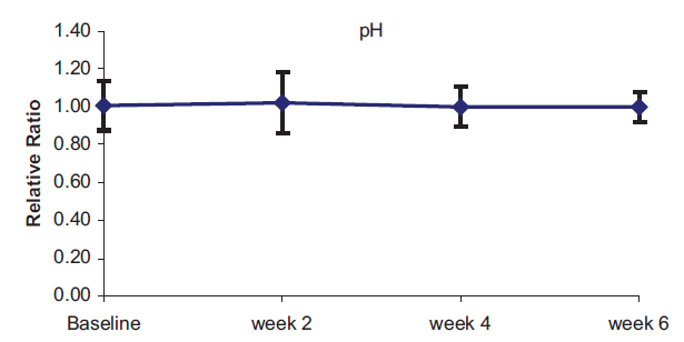
Figure 8. Changes of pH over time. Data are shown as mean ± SD and are expressed as the ratios of the laser-treated areas to the control areas. There was no statistically significant difference between the areas.
Discussion
The efficacy of laser treatment for hair removal is superior to conventional treatments such as shaving, wax epilation and electrolysis, although there is no evidence for a complete and persistent hair removal efficacy. In an intervention review, it was shown that diode lasers had a short-term effect of approximately 50% hair reduction within up to 6 months after the treatment. In addition, in a meta-analysis, it was shown that mean hair reduction at least 6 months after the last treatment was 57.5% after three sessions with the diode laser.
The specific chromophore for laser hair removal is melanin, which absorbs the light emitted by the laser at a specific wavelength. Depending on the amount of energy received by the follicle during the laser treatments, the final result may be the swelling of keratinocytes, scattered apoptotic/necrotic keratinocytes, or full-thickness necrosis of the follicle. Following these early period changes, the number of catagen hairs increases due to early change from the anagen to catagen phase, or hair may completely drop out of follicles. During the period after the catagen/telogen phase, it is possible that the damaged follicles return to the anagen phase, which then would result in regrowth of some of the unwanted hairs. However, a progressive decrease in hair counts is observed after laser treatments, which is probably due to the fact that a significant number of the follicles which are in the catagen phase cannot restart the cycle. In a clinical study which seems to verify the above information, regrowth of the hairs, depending on the fluence used, ranged from 22% to 31% during the 1-month follow-up after a single treatment performed with the diode laser. It then remained stable between 65% and 75% during the average follow-up of 20 months. We evaluated only short-term changes of hair growth in our study and hair growth with the diode laser was determined as 49.68% in 2 weeks, 46.01% in 4 weeks, and 48.15% in 6 weeks. The difference between hair growth in the first month in our study and hair growth in Lou`s study may be due to individual differences between patients, differences in application areas and differences in the laser parameters.
Although the purpose of laser hair removal is selective damage to the follicles, the thermal injury likely to develop can cause damage in appendageal structures and epidermis as well. It revealed that there was vacuolization and swelling of basal keratinocytes in the overlying epidermis in addition to damage to hair follicles in one single specimen after diode laser treatment. In addition, the researcher demonstrated that there was an apparent decrease in sebaceous gland size after ruby laser treatment due to the possible thermal effects of laser since human sebaceous glands contain no pigmented melanocytes. We postulate that these existing limitations on tissue selectivity can cause changes to the biophysical properties of skin. Previous studies demonstrated that regional and seasonal changes and age-related variation have effects on biophysical parameters of the skin. However, our trial was designed as a right – left controlled study to exclude possible confounders.
Transient erythema is mainly caused by the heat resulting from the absorption of laser light by epidermal melanin and is not always recognized as an adverse event. The thermal heat generated by laser – skin interaction results in vasodilation, caused by the release of prostaglandin and direct thermal damage of vessels. Erythema normally clears within several hours with adequate cooling, but it has been reported to last as long as 7 days. In our study, a statistically significant increase was determined in EI in the second week in the laser-treated area compared to the control area, which shows that the erythema response with the diode laser can continue up to the second week. Although pigmentary changes caused by the absorption of laser energy by epidermal melanin are among the most common side effects of non-ablative laser therapies. There was no difference in MI with the diode laser in our study. In addition, we did not detect any changes in the biophysical properties of skin, including transepidermal water loss, the capacity of stratum corneum hydration, sebum and pH level. As a result, our findings show that the diode laser can perform a significant reduction in the hair amount without significant epidermal damage, at least for a short period.
Welcome to visit our Website to know more information aboout the Professional aesthetic equipment
LET'S GET IN TOUCH

Privacy statement: Your privacy is very important to Us. Our company promises not to disclose your personal information to any external company with out your explicit permission.

Fill in more information so that we can get in touch with you faster
Privacy statement: Your privacy is very important to Us. Our company promises not to disclose your personal information to any external company with out your explicit permission.